Kondapur & Secunderabad

Cone Beam Computed Tomography

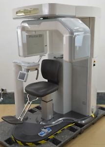
Cbct is a medical imaging technique consisting of X-ray computed tomography where the X-rays are divergent, forming a cone.

Population Inversion

This occurs when the two sources are mixed together and photons are given off thereby producing the “LASER” activity is resonated or reflected back and forth within an optical resonator and amplified. A portion of this activity is then emitted as a LASER beam. Beam splitters or mirrors are used to capture the particular portion of the LASER beam that is needed to perform the surgical operation.


A CT image comprises slices along a plane of the object being viewed. The slices, called voxels, are then reconstructed. The x-ray energy from a CT scan is directed toward an object from multiple orientations, and the decrease in intensity is measured along a series of linear paths In layman’s terms,CBCT is a compact, faster and safer version of the regular CT. Through the use of a cone shaped X-Ray beam, the size of the scanner, radiation dosage and time needed for scanning are all dramatically reduced.


A typical CBCT scanner can fit easily into any dental practice and is easily accessible by patients. The time needed for a full scan is typically under one minute and the radiation dosage is up to a hundred times less than that of a regular CT scanner.

Indications of CBCT in the Maxillofacial Region

  • Evaluation of the jaw bones to assess the feasibility of placing dental implants at specific sites in the jaws. This ensures that every possible precaution has been made to reduce the risk of involvement of the nerves in the lower jaw, and the sinuses and nose in the upper jaw.
  • Evaluation of the status of previously placed implants.
  • Evaluation of the hard tissue (bones) of the tempro-mandibular joint (TMJ).
  • Evaluation of abnormalities (pathology) in or affecting the bonesEvaluate extent of alveolar ridge resorption.
  • Assessment of relevant structures prior to orthodontic treatment such as the presence and position of impacted canine and third molar teeth.
  • Assessing symmetry of the face (cephalometrics)
  • To permit 3D reconstructions of the bones or the fabrication of a Biomodel of the face and jaws
  • Assessing the mandibular nerve prior to the removal of impacted teeth, especially the lower wisdom teeth
  • A CBCT scan provides precise information about pathology, the locations and dimensions of vital anatomical structures, bone quality, and any issues that might interfere with treatment planning.

Specific Applications

Benefits:

  • X-Ray Radiation exposure to the patient is up 10 times less than a regular CT scanner.
  • Much faster scan time. Scan on a CBCT takes between 10-40 sec, while on a regular CT scanner it takes a few minutes.
  • Unlike regular x-rays CT scans can discriminate between many types of tissue including bone, teeth, nerves and soft tissue.
  • CT scans are noninvasive, and can eliminate the need for exploratory surgery in some cases.
  • CT can identify the effects of conditions such as infection and tumors.
  • A cost effective tool for imaging a wide range of clinical problems.

A periapical X-ray is a specific type of intraoral X-ray that is used to investigate the structural integrity of an individual tooth. A periapical X-ray provides an image of a tooth from the tooth’s crown to the tip of its root. Periapical X-rays provide a more highly focused, finely detailed image than common bitewing X-rays that survey three-to-four teeth at a time.

RVG – digital radiovisiography

RVG – digital radiovisiography is the latest X-raytechnology in dentistry. Digital radiovisograph is equipped with special image analysis software, and in digital technology it is possible to enlarge the images and their saving in the patient’s file.

Some of the advantages of the digital radiovisiography

  • Reduced radiation of the patient compared with classic X-ray up to 80%
  • Faster imaging without X-ray film and developing images
  • Digital intraoral sensor is used instead of X-ray film
  • Immediate imaging on the computer screen
  • high quality of the digital image that can be analyzed and processed
  • saving images in the patient’s file
  • children friendly for reduced radiation, if imaging is necessary

Orthopantomogram (OPG)

An Orthopantomogram (OPG), also known as an “Orthopantogram” is a panoramic scanning dental X-ray of the upper and lower jaw. It shows a two-dimensional view of a half-circle from ear to ear.

OPGs are used by Dentists to provide information on

  • Impacted wisdom teeth
  • Periodontal bone loss
  • Finding the source of dental pain
  • Assessment for the placement of dental implants
  • Orthodontic assessment

Need More Info about CBCT Imaging


CALL US FOR A QUICK APPOINTMENT
+91-9963100600, +91-9640100600

KIMS Dental Care provide Cbct imaging service which is a faster and safer version of the regular CT.
Kondapur Branch
  • Mon-Sat
    09:00 AM - 09:00 PM
    Sunday
    10:00 AM - 02:00 PM
Contact Info
Secunderabad Branch
  • Mon-Sat
    09:00 AM - 07:00 PM
    Sunday
    Only on Call
Contact Info
Ask the Experts

Your message was sent successfully!

Something went wrong, try refreshing and submitting the form again.


Specialist Dental Practice

Our dentists are committed to improving your general dental health and restoring your smile. We are a specialist dental practice also offering the best and most affordable dental implants for your oral health and rehabilitation. Make a FREE dental implants consultation and assessment for a personal treatment plan and meet our friendly and professional staff.


Kondapur Location
  • KIMS Dental Care
    # 1-112 / 86, Survey No 55/ EE,
    Kondapur, Serilingampally Mandal,
    Hyderabad - 500084
Secunderabad Location
  • KIMS Dental Care
    1-8-31/1, Minister Road,
    Secunderabad - 500003

Kondapur


Secunderabad